5-ALA Fluorescence Guides Neurosurgeons for More Complete Resections
July 7, 2021Categories: Cover Story
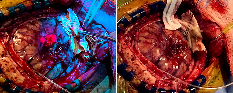 A comparison of a high-grade glioma shows how cancerous cells take on a luminescent pink color under 5-ALA florescent-guided resection (left) versus conventional surgery. Photo: NX Development Corp.
Neurosurgeons Ammar Hawasli, MD, PhD, and Stephen Reintjes Jr., MD, with Meritas Health Neurosurgery, are now using a progressive treatment that allows for the more precise removal of malignant gliomas and glioblastomas. With 5-aminolevulinic acid-guided tumor resection, cancerous cells fluoresce pink under ultraviolet light, while healthy tissue glows blue.
These tumors inherently possess highly invasive borders that are difficult to distinguish from normal brain tissue. This can make traditional surgical resection by intraoperative microscope incomplete because they are not visible on MRI and therefore inoperable.
“In the past, these tumors were removed with the aid of intraoperative MRI, where a surgeon could assess the extent of resection in the OR,” Dr. Hawasli said. “5-ALA replaces intraoperative MRI because we can see the tumor cells that cannot be seen under regular microscopic white light. With this advanced treatment, patients need not go to a tertiary care center. They can stay here and know they are receiving state-of-the art brain tumor surgery.”
Pink Pathway
A few hours before surgery, patients drink a small vial of 5-ALA, which is a natural compound. Malignant cells uptake and break down the 5-ALA, which then metabolizes into fluorescent pink protoporphyrin IX under blue light.
“We use 5-ALA for any malignant brain tumor, and there are no size restrictions,” Dr. Hawasli said.
Because the drug is phototoxic, patients are kept under low light for about 48 hours after surgery. Their length of stay averages two to five days.
Dr. Hawasli trained on the surgery during his residency in 2015 in Ireland. In Europe, the surgery has long been a standard of care. Before joining Meritas Health Neurosurgery, he performed 5-ALA surgeries in clinical trials in his previous role at Washington University.
Dr. Reintjes Jr., who has trained in its use, also will utilize this technology at North Kansas City Hospital.
The Food and Drug Administration approved the surgery in 2017. In 2019, a neurosurgeon at University Hospitals Cleveland Medical Center performed the first U.S. surgery.
Quality of Life
In a study published in the May 2006 issue of Lancet Oncology, surgeons achieved greater resection with 5-ALA over white-light microscopy. In “Fluorescence-Guided Surgery With 5-Aminolevulinic Acid for Resection of Malignant Gliomas: A Randomized Controlled Multicenter Phase III Trial,” researchers found a 65% rate of complete resection in the 5-ALA group, compared to 35% in the white-light microscopy group. The study involved 322 patients ages 23-73 with suspected malignant gliomas.
“We know the extent of resection, especially with high-grade gliomas and glioblastomas, correlates with a patient’s longevity,” Dr. Hawasli added. “Ten years ago, clinical trials gave people with a glioblastoma one year to live. With 5-ALA guided surgery and other treatments, patients enjoy a much longer life. My patients who have had high-grade gliomas know if they recur, there is technology available to help them live longer. They are excited that things are improving.”
Ammar Hawasli, MD, PhD
Dr. Hawasli earned his combined medical and doctorate in philosophy degrees from the University of Texas Southwestern Medical Center. He was a neurological surgery resident and chief resident and then a fellow in complex spine surgery at Washington University School of Medicine, Barnes-Jewish Hospital and St. Louis Children’s Hospital. During his residency, he was a specialist neurosurgery registrar (resident) at Beaumont Hospital in Dublin, Ireland, where he used 5-ALA routinely.